Micro-CT
Computed tomography (CT) is used as a non-destructive technique to visualize and study external and internal structures of objects. In investigating rare fossil material, this method is state-of-the-art. By combining micro-CT scanning with different techniques of staining, also soft tissue, e.g. muscles and tendons, can be visualized non-invasively. Researchers from the University of Vienna and other research institutions as well as curators of natural history museums apply this technique to realize a wide variety of research and conservation objectives. It is a multipurpose technique to allow in-depth investigation of objects without destroying the investigated material.
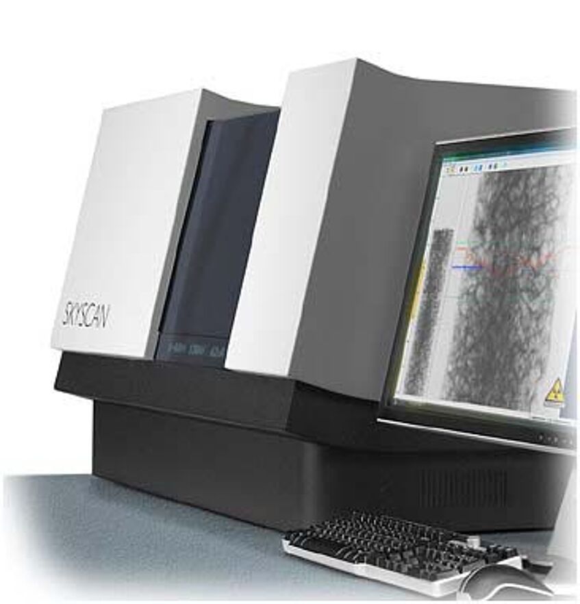
The Department of Palaeontology at the University of Vienna holds a state-of-the-art reflection micro-CT.
Micro-CT
In December 2010 the Department of Palaeontology acquired a Skyscan 1173 Desktop-Micro-CT (Bruker), which is a high energy spiral scan micro-CT for analysing both dense and low dense materials. It includes a 130kV microfocus X-ray source with improved stability of focal spot position, a large format (>5Mp) flat panel sensor with special protection by lead-glass fiber-optic window for long lifetime under high-energy X-ray and a precision object manipulator for large (up to 140mm) and heavy (several kg) objects with embedded micropositioning stage.
Micro-CT team
Univ.-Prof. Dr. Jürgen Kriwet - Head of micro-CT Facility
Dr. Cathrin Pfaff - Micro-CT Specialist
Specifications are:
- X-ray source: 40-130kV, 8W, <5µm spot size
- X-ray detector: distortion-free Flat Panel sensor 2240x2240 pixels, 12-bit
- Maximum object size: 40mm in diameter, 200mm in length (100-140mm scanning length)
- Spatial resolution : <4-5µm detail detectability (depending on sample size), 7-8µm low-contrast resolution
- Reconstruction: single PC or cluster volumetric reconstruction (Feldkamp algorithm)
- Radiation safety: <1µSv / h at any point on the instrument surface
Basic principles
The process of micro-computer tomography includes:
- Scanning of object
- Reconstruction
- Visualisation
- Data collection and storage
During scanning 2D representations of an object based on material density measured by X-ray transmission is produced. Slices are then stacked by using different programs of virtual reconstruction to produce a virtual 3D cast by assigning CT (grey) values to each voxel (3D pixels of 2D slice). The resulting 3D image is a mathematical depiction of the object and thus is suitable for quantitative analyses.
Scanning requests, costs and protocol
- Download and complete the micro-CT scanning request for applying for scanning time.
- Email complete form to Cathrin Pfaff.
- Duration and costs of scanning are based on specimen size, specimen dimensions, special requests
concerning the field or region of interest, material, object preservation, measurement and analysis,
required format, etc.
- Objects must be stored in a sealed box or airtight bags or boxes for wet specimens.
- Specimen mounts are provided by the facility.
- Slices can be exported in a wide variety of formats and can be collected using hard drives (NTFS format).
- External hard drives must be formatted prior to use and all hard drives, cables, etc. must be labelled with
user's name.
- Contact Cathrin Pfaff & Jürgen Kriwet to discuss your project and costs if necessary.
TESCAN Vega 4 GMU-Rasterelektronenmikroskop

Copyright C.H. Baal
Das Rasterelektronenmikroskop des Typs TESCAN Vega 4 GMU wird vom Institut für Paläontologie in Zusammenarbeit mit den erdwissenschaftlichen Subeinheiten der FGGA betrieben.
Die Einrichtung für Rasterelektronenmikroskopie und Ionenstrahlanwendungen umfasst ein konventionelles Wolframfilament-Rasterelektronenmikroskop (TESCAN Vega 4 GMU) und ein Feldemissions-Rasterelektronenmikroskop mit fokussiertem Ionenstrahl (FEI Quanta 3D FEG). Die REM-Einrichtungen werden von den erdwissenschaftlichen Subeinheiten der Fakultät für Geowissenschaften, Geographie und Astronomie gemeinsam genutzt.
Das TESCAN Vega 4 GMU ist unser wesentliches Werkzeug für standardmäßige Elektronenmikroskopie, welches eine schnelle Charakterisierung von Mineralen, Fossilien und Gesteinsoberflächen erlaubt. Das Gerät ist mit Sekundärelektronen-Detektoren für den Einsatz im Hoch- und Niedrigvakuum ausgestattet, sowie mit einem Rückstreuelektronen-Detektor, einem energiedispersiven Röntgenspektrometer für semi-quantitative mikrochemische Analysen (Oxford AztecLiveLite EDS) und einem Kathodolumineszenz-Detektor (TESCAN CL).
Ansprechperson für weitere Informationen zu dem TESCAN Vega 4 GMU:
Nutzungsordnung SEM-Labor
Dateigröße: 660 kB
Spezifikationen des TESCAN Vega 4 GMU
Dateigröße: 449 kB